


Global obesity trials are scaling fast with GLP‑1 therapies, making complete data capture essential to preserve scientific integrity and timelines. ICON implemented a pragmatic DXA strategy—modified hand positioning and a dual‑scan workflow with centralized review—that preserved full visceral fat datasets, preventing “silent attrition” from incomplete imaging.
Key takeaways
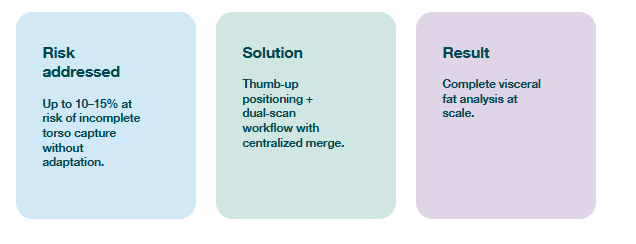
- Up to 10–15% of participants can fall outside the standard DXA field‑of‑view, jeopardizing visceral fat calculations.
- Two simple, scalable techniques (thumb‑up hand positioning and dual‑scan + centralized merge) enabled complete datasets across a 100‑site global network.
- Full data capture strengthens integrity, timelines, and budgets in competitive GLP‑1 obesity trials.
Context — GLP‑1 scale raises the stakes for complete datasets
Obesity trials are scaling rapidly. With the rise of GLP-1 therapies like semaglutide and tirzepatide, sponsors are launching global studies with thousands of participants across hundreds of sites, all competing for overlapping target populations. Eli Lilly’s SURMOUNT-1 study, for example, enrolled over 2,500 patients to demonstrate the efficacy of tirzepatide, while Novo Nordisk’s semaglutide trial, STEP 1, enrolled nearly 2,000 patients and both companies included a whole-body DXA imaging sub study to assess body composition.
This surge in clinical trial activity within obesity, especially around GLP-1 therapies, has created intense competition for eligible participants. Sponsors are under more pressure to recruit faster, retain better and maximise data capture from every individual enrolled to deliver robust, quality data and reach market ahead of the competition. When approximately 10-15% of carefully recruited and engaged participants are at risk of being lost due to logistical imaging challenges, imaging becomes more than a protocol requirement—it can be leveraged as a parallel retention strategy to ensure 100% data capture.
The imaging gap: A hidden risk to retention
DXA scans are widely used in obesity trials for their speed, comfort and ability to differentiate between fat, bone and lean mass. They are particularly valuable for identifying visceral fat, a key indicator of cardiometabolic risk. However, unlike MRI, DXA machines are not designed to accommodate larger body sizes. For up to 15% of participants, the torso data may not be fully captured using standard DXA protocols, representing a significant loss of participant data.
The standard protocol is to image participants on an offset, which for most individuals would capture the full torso and full limbs on the offset side. The data for the torso would be captured and the data for the limbs would then be mirrored to provide an approximate dataset for both sides of the body. For the 10-15% of individuals whose torso would be too large to fit within the imaging field, this means the DXA machine would be unable to make the appropriate visceral fat calculations.
This limitation posed a risk not only to data integrity but to functional participant retention. Individuals who undergo screening and imaging but yield incomplete data may be excluded from analysis, effectively lost to the study despite being enrolled.
ICON’s small but mighty imaging solution for 100% data capture
ICON recognised the increasing risk of participant data collection tied to the logistics and limitations of the DXA machines themselves. To avoid any data loss, ICON implemented a scalable imaging strategy that enabled full data capture by making small adjustments to scan techniques.
Two tailored techniques were introduced:
- Modified hand positioning: Adjusting the hand to a thumb-up position created additional space within the imaging field, improving torso coverage.
- Dual-scan method: Sites acquired two scans, when necessary, one offset for limb data and one centred for full abdominal imaging. These were combined via centralised automation to produce a complete dataset.
To ensure consistency across the global network of 100 sites for this particular trial, ICON’s Medical Imaging and Cardiac Safety (IMC) team provided detailed imaging manuals and training modules. Imaging data was centralised and reviewed by specialists who analysed, validated and merged the scans, preserving both accuracy and operational efficiency.
Future-proofing obesity imaging
As obesity trials evolve to assess not just weight loss but the quality of that loss by distinguishing fat from muscle and subcutaneous from visceral fat, imaging precision becomes even more critical in understanding the long-term impacts of these therapies. While MRI has more robust fat and muscle discernment capabilities—quantifying subcutaneous, intramuscular and visceral fats with the ability to exclude organs and major blood vessels from muscle tissue, DXA provides quick and efficient scans that quantify the torso, limbs and head and can separate visceral fat from subcutaneous fat within the abdomen. Identifying visceral fat is important as it has a strong, positive association with major adverse cardiovascular events (MACE). These distinctions and the innovations they support are only possible with high-quality imaging.
DXA remains the most feasible modality for large-scale trials, offering speed and cost efficiency while fulfilling key measurement needs. MRI, while more precise, is often reserved for smaller subgroups or early-phase studies due to its higher cost and complexity.
ICON’s approach ensures that DXA can be implemented effectively even in diverse populations, avoiding the silent attrition of participants whose scans would otherwise be incomplete. This strategy complements the multi-level, multi-channel recruitment and engagement approaches used in obesity trials to combat high dropout rates, supporting trial timelines and data integrity.
Outcome: Complete data, full value
By enabling complete visceral fat analysis for every participant with a scalable DXA scan technique, ICON is helping sponsors avoid data loss from an estimated 10–15% of their cohorts. This not only preserves the scientific integrity of their trials but also protects their operational timelines and budgets.
In today’s competitive obesity trial landscape, where recruitment is hard-won and retention is paramount, proactive imaging design is a strategic advantage. Connect with us to learn how ICON’s Medical Imaging team can maximise your data capture to ensure the high data integrity and retention for meaningful outcomes.
To learn more about how obesity trials are scaling with GLP‑1 therapies download the following document
